Comparison of optical coherence tomography measurements between high hyperopic and low hyperopic children
| dc.contributor.author | Dikkaya, Funda | |
| dc.contributor.author | Karaman Erdur, Sevil | |
| dc.date.accessioned | 2020-04-27T05:56:41Z | |
| dc.date.available | 2020-04-27T05:56:41Z | |
| dc.date.issued | 2020 | en_US |
| dc.identifier.citation | Dikkaya, F. ve Karaman Erdur, S. (2020). Comparison of optical coherence tomography measurements between high hyperopic and low hyperopic children. Therapeutic Advances in Ophthalmology, 12. https://dx.doi.org/10.1177/2515841419899819 | en_US |
| dc.identifier.issn | 2515-8414 | |
| dc.identifier.uri | https://hdl.handle.net/20.500.12511/5202 | |
| dc.identifier.uri | https://dx.doi.org/10.1177/2515841419899819 | |
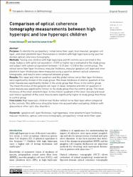
| dc.description.abstract | Purpose: To identify the peripapillary retinal nerve fiber layer, total macular, ganglion celllayer, and inner plexiform layer thicknesses in children with high hyperopia using spectraldomain optical coherence tomography.Methods: Twenty-one children with high hyperopia and 20 controls were enrolled in thisstudy. Subjects with spherical equivalent +5.0D or higher were evaluated in the study groupand subject with spherical equivalent between +0.25 and +2.0 D in the control group. Theretinal nerve fiber layer thickness, macular thickness, macular ganglion cell layer and innerplexiform layer thicknesses were measured using a spectral domain optical coherencetomography, and results were compared between groups.Results: The nasal and inferior quadrant and the global retinal nerve fiber layer thicknesswere significantly thicker in the study group. The mean thickness of inferior quadrant of theinner macula was significantly thicker in the study group than those in the control group.The mean thickness of the ganglion cell layer in nasal, temporal and inferior quadrant ofouter macula was significantly thinner in the study group than the control group. The meanthickness of the inner plexiform layer in the inferior quadrant of the inner macula and nasaland inferior quadrant of the outer macula were significantly higher in study group than thosein control group.Conclusion: High hyperopic children had thicker retinal nerve fiber layer when comparedto the controls. This difference should be taken into account when evaluating children withglaucoma or other optic disc disorders. | en_US |
| dc.language.iso | eng | en_US |
| dc.publisher | SAGE Publications Ltd | en_US |
| dc.rights | info:eu-repo/semantics/openAccess | en_US |
| dc.rights | Attribution-NonCommercial 4.0 International | * |
| dc.rights.uri | https://creativecommons.org/licenses/by-nc/4.0/ | * |
| dc.subject | Ganglion Cell Layer Thickness | en_US |
| dc.subject | High Hyperopia | en_US |
| dc.subject | Inner Plexiform Layer Thickness | en_US |
| dc.subject | Macular Thickness | en_US |
| dc.subject | Optical Coherence Tomography | en_US |
| dc.subject | Peripapillary Retinal Nerve Fiber Layer | en_US |
| dc.title | Comparison of optical coherence tomography measurements between high hyperopic and low hyperopic children | en_US |
| dc.type | article | en_US |
| dc.relation.ispartof | Therapeutic Advances in Ophthalmology | en_US |
| dc.department | İstanbul Medipol Üniversitesi, Tıp Fakültesi, Cerrahi Tıp Bilimleri Bölümü, Göz Hastalıkları Ana Bilim Dalı | en_US |
| dc.authorid | 0000-0003-2312-2521 | en_US |
| dc.authorid | 0000-0001-9829-7268 | en_US |
| dc.identifier.volume | 12 | en_US |
| dc.relation.publicationcategory | Makale - Uluslararası Hakemli Dergi - Kurum Öğretim Elemanı | en_US |
| dc.identifier.doi | 10.1177/2515841419899819 | en_US |
Bu öğenin dosyaları:

Bu öğe aşağıdaki koleksiyon(lar)da görünmektedir.
-
Makale Koleksiyonu [3777]
Article Collection -
PubMed İndeksli Yayınlar Koleksiyonu [4230]
PubMed Indexed Publications Collection -
WoS İndeksli Yayınlar Koleksiyonu [6631]
WoS Indexed Publications Collection

Aksi belirtilmediği sürece bu öğenin lisansı: info:eu-repo/semantics/openAccess


