| dc.contributor.author | Kırgız, Ahmet | |
| dc.contributor.author | Karaman Erdur, Sevil | |
| dc.contributor.author | Şerefoğlu Çabuk, Kübra | |
| dc.contributor.author | Atalay, Kürşat | |
| dc.contributor.author | Aşık Nacaroğlu, Şenay | |
| dc.date.accessioned | 2020-03-04T06:41:50Z | |
| dc.date.available | 2020-03-04T06:41:50Z | |
| dc.date.issued | 2019 | en_US |
| dc.identifier.citation | Kırgız, A., Karaman Erdur, S., Şerefoğlu Çabuk, K., Atalay, K. ve Aşık Nacaroğlu, Ş. (2019). Alterations in corneal biomechanical and topographic features after accelerated crosslinking: 1-year results. Beyoglu Eye Journal, 4(2), 108-114. http://doi.org/10.14744/bej.2019.44154 | en_US |
| dc.identifier.issn | 2459-1777 | |
| dc.identifier.issn | 2587-0394 | en_US |
| dc.identifier.uri | http://doi.org/10.14744/bej.2019.44154 | |
| dc.identifier.uri | https://hdl.handle.net/20.500.12511/4975 | |
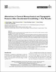
| dc.description.abstract | Objectives: To determine the biomechanical and topographic alterations within the first year after accelerated crosslink-ing (CXL) treatment in patients with keratoconus. Methods: In this prospective study, 52 eyes of 52 patients with progressive keratoconus underwent accelerated CXL were included. All patients had a detailed preoperative ophthalmologic examination, including slit-lamp evaluation, Gold-mann tonometry, fundoscopy, topography by Scheimpflug imaging (Sirius), and corneal biomechanical evaluation with a biomechanical waveform analysis device (ORA). Alterations in visual acuity and topographic findings were evaluated before the treatment and at 12 months follow-up. Corneal biomechanical features were obtained before the treatment, and at 1st, 3rd, 6th and 12th months. Results: Uncorrected-visual acuity and best-corrected visual acuity both statistically significantly improved at 12th month (p=0.001). There were no statistically significant differences in keratometry values, whereas maximum K (AKfront) and symmetry index front (SIfront) decreased significantly (p=0.015 and p=0.009, respectively). Corneal thinnest point and volume also decreased significantly at 12th month (p=0.001 for both). Goldmann-correlated intraocular pressure (IOPg) and corneal compensated IOP (IOPcc) values transiently increased in the first three months, while corneal hysteresis (CH) and the corneal resistance factor (CRF) transiently decreased, with the difference not statistically significant (p>0.05). However, central corneal thickness significantly decreased at the end of the 12th month (p=0.001). Conclusion: Accelerated CXL seems to be effective in stopping the progression of keratoconus. Our findings showed transient alterations in biomechanical features, which will end with the preoperative values at the end of the 12th month. Further studies are needed to demonstrate the changes in corneal biomechanics in vivo. | en_US |
| dc.language.iso | eng | en_US |
| dc.rights | info:eu-repo/semantics/openAccess | en_US |
| dc.rights | Attribution-NonCommercial 4.0 International | * |
| dc.rights.uri | https://creativecommons.org/licenses/by-nc/4.0/ | * |
| dc.subject | Accelerated Crosslinking | en_US |
| dc.subject | Corneal Biomechanics | en_US |
| dc.subject | Corneal Topography | en_US |
| dc.subject | Corneal Hysteresis | en_US |
| dc.subject | Keratoconus | en_US |
| dc.title | Alterations in corneal biomechanical and topographic features after accelerated crosslinking: 1-year results | en_US |
| dc.type | article | en_US |
| dc.relation.ispartof | Beyoglu Eye Journal | en_US |
| dc.department | İstanbul Medipol Üniversitesi, Tıp Fakültesi, Cerrahi Tıp Bilimleri Bölümü, Göz Hastalıkları Ana Bilim Dalı | en_US |
| dc.authorid | 0000-0001-9829-7268 | en_US |
| dc.identifier.volume | 4 | en_US |
| dc.identifier.issue | 2 | en_US |
| dc.identifier.startpage | 108 | en_US |
| dc.identifier.endpage | 114 | en_US |
| dc.relation.publicationcategory | Makale - Ulusal Hakemli Dergi - Kurum Öğretim Elemanı | en_US |
| dc.identifier.doi | 10.14744/bej.2019.44154 | en_US |


