| dc.contributor.author | Yalçın Özbek, Merve | |
| dc.contributor.author | Aydın, Rukiye | |
| dc.contributor.author | Özsütçü, Mustafa | |
| dc.contributor.author | Kocabora, Mehmet Selim | |
| dc.date.accessioned | 08.07.201910:49:13 | |
| dc.date.accessioned | 2019-07-08T20:18:30Z | |
| dc.date.available | 08.07.201910:49:13 | |
| dc.date.available | 2019-07-08T20:18:30Z | |
| dc.date.issued | 2017 | en_US |
| dc.identifier.citation | Yalçın Özbek, M., Aydın, R., Özsütçü, M. ve Kocabora, M. S. (2017). Asimetrik retinitis pigmentosa. MN Oftalmoloji, 24(4), 245-247. | en_US |
| dc.identifier.issn | 1300-4786 | |
| dc.identifier.uri | https://hdl.handle.net/20.500.12511/321 | |
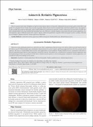
| dc.description.abstract | Otuz iki yaşında kadın hasta kliniğimize sol gözde bulanık görme şikayeti ile başvurdu. Oftalmolojik muayenesinde, görme keskinlikleri sağgözde 1,0, sol gözde 0,8 idi. Fundus muayenesinde sağ gözde oldukça küçük sınırlı bir alanda birkaç adet kemik spikülü benzeri hiperpigmente alan görüldü. Sol gözde her kadranda, yaygın kemik spikülü tarzı pigmentasyon saptandı. Fundus otofloresans incelemesinde sol gözde maküla etrafında dikkat çeken hiperreflektif halo görünümü mevcuttu. Muayene ve fundus bulguları sonucunda tek taraflı retinitis pigmentosa olarak düşünülen olguya elektrofizyolojik testler yapıldı. Yapılan elektroretinografisinde sol gözde elektroretinografi yanıtları azalmıştı. Hastadamevcut bulgular ışığında asimetrik retinitis pigmentosa düşünüldü. | en_US |
| dc.description.abstract | Thirty-two-years-old female patient was referred to our clinic complaining of blurred vision in her left eye. Best-corrected visual acuity in her left eye was 0.8. Dilated fundoscopic examination demonstrated a few pieces of quite small hyperpigmented areas like bone spicules in the right eye. The left eye revealed diffuse bone spicules style pigmentation in each quadrant. Fundus autofluorescence examination of the left eye showed remarkable hyperreflective halo appearance around the macula. The patient was considered to be unilateral retinitis pigmentosa and electrophysiological tests were performed. At the left eye had decreased responses in electroretinography. Asymmetric retinitis pigmentosa was considered with present findings at the patient. | en_US |
| dc.language.iso | tur | en_US |
| dc.rights | info:eu-repo/semantics/openAccess | en_US |
| dc.subject | Asimetrik Retinitis Pigmentosa | en_US |
| dc.subject | Elektroretinografi | en_US |
| dc.subject | Fundusotofloresans | en_US |
| dc.subject | Retinal Dejenerasyon | en_US |
| dc.subject | Asymmetric Retinitis Pigmentosa | en_US |
| dc.subject | Electroretinography | en_US |
| dc.subject | Fundus Autofluorescence | en_US |
| dc.subject | Retinal Degeneration | en_US |
| dc.title | Asimetrik retinitis pigmentosa | en_US |
| dc.title.alternative | Asymmetric retinitis pigmentosa | en_US |
| dc.type | other | en_US |
| dc.relation.ispartof | MN Oftalmoloji | en_US |
| dc.department | İstanbul Medipol Üniversitesi, Tıp Fakültesi, Cerrahi Tıp Bilimleri Bölümü, Göz Hastalıkları Ana Bilim Dalı | en_US |
| dc.authorid | 0000-0003-0668-3749 | en_US |
| dc.authorid | 0000-0001-8954-5055 | en_US |
| dc.authorid | 0000-0001-5335-3860 | en_US |
| dc.identifier.volume | 24 | en_US |
| dc.identifier.issue | 4 | en_US |
| dc.identifier.startpage | 245 | en_US |
| dc.identifier.endpage | 247 | en_US |
| dc.relation.publicationcategory | Diğer | en_US |