Unusual cause of chest pain mimicking acute myocardial infarction: Congenital left ventricular aneurysm
Citation
Karaca, O., Kayhan, B., Omaygenç, O., Çakal, B. ve Türkoǧlu, H. (2015). Unusual cause of chest pain mimicking acute myocardial infarction: Congenital left ventricular aneurysm. Journal of Clinical and Diagnostic Research, 9(1), OJ01-OJ02. https://dx.doi.org/10.7860/JCDR/2015/11329.5432Abstract
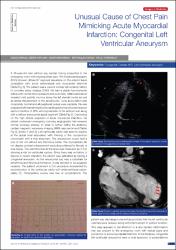
A 36-year-old man without any cardiac history presented to the emergency room with ongoing chest pain. The Electrocardiography (ECG) showed diffuse ST segment elevations on the anterior leads compatible with acute anterolateral wall myocardial infarction [Table/Fig-1]. The patient was a current smoker with a family history of coronary artery disease (CAD). He had a stable hemodynamic status with normal blood pressure and pulse rate. Initial examination revealed mild systolic murmur along the left sternal border as well as lateral displacement of the apical pulse. Lung auscultation was completely normal and all peripheral pulses were palpable. He was evaluated with transthoracic echocardiography that showed reduced ejection fraction of 38% with hypokinesis of the anterior wall along with a diffuse aneurysmal apical segment [Table/Fig-2]. Depending on the high clinical suspicion of acute myocardial infarction, the patient underwent emergency coronary angiography that revealed normal coronary arteries. In order to further define the anatomy, cardiac magnetic resonance imaging (MRI) was performed [Table/ Fig-3], [Video-1 and 2]. Left ventricular cavity was seen to expand at the apical level associated with thinning of the myocardium concordant with a true aneurysm. The aneurysmal pouch had a size of 4x5 cm without any thrombus inside.
xmlui.dri2xhtml.METS-1.0.item-scopusquality
Q3Source
Journal of Clinical and Diagnostic ResearchVolume
9Issue
1Collections
- Makale Koleksiyonu [3770]
- PubMed İndeksli Yayınlar Koleksiyonu [4224]
- Scopus İndeksli Yayınlar Koleksiyonu [6561]
- WoS İndeksli Yayınlar Koleksiyonu [6621]


