A pictorial view to tarsal coalition: The presentation of two children with foot pain
Künye
Örmeci, T., Kılıçarslan, R., Durmuş, O., Malkoç, M. ve Çakar, E. (2014). A pictorial view to tarsal coalition: The presentation of two children with foot pain. Acta Reumatologica Portuguesa, 39(4), 347-348.Özet
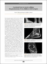
Two male patients aged 9 and 16 years were admitted with complaints of ankle pain and intoeing. There was no history of trauma and arthritis. In the first case, radiography examination revealed a “C sign” (a cortical continuity from the medial portion of the talus to the sustentaculum tali), a narrowing in the middle subtalar joint space, and convexity in the lower margin of the sustentaculum tali (Figure 1). Magnetic resonance imaging (MRI) showed triple coalition in the anterior subtalar joint (talocalcaneonavicular joint) in the first case (Figures 2 and 3) and fibrocartilaginous coalition at the posterior subtalar joint in the second case (Figure 4). Narrowing in the joint spaces, findings of early degeneration, and medullary edema in the adjacent bone were observed at the sites of coalition (Figures 3 and 4). Nonoperative treatment, such as physical therapy, exercises, and analgesic medication were applied.
WoS Q Kategorisi
Q4Scopus Q Kategorisi
Q3Kaynak
Acta Reumatologica PortuguesaCilt
39Sayı
4Koleksiyonlar
- Makale Koleksiyonu [3672]
- PubMed İndeksli Yayınlar Koleksiyonu [4080]
- Scopus İndeksli Yayınlar Koleksiyonu [6346]
- WoS İndeksli Yayınlar Koleksiyonu [6465]


