| dc.contributor.author | Karaarslan, Numan | |
| dc.contributor.author | Yılmaz, İbrahim | |
| dc.contributor.author | Yaşar Şirin, Duygu | |
| dc.contributor.author | Baykız, Derya | |
| dc.contributor.author | Demirkıran, Aykut | |
| dc.contributor.author | Ateş, Özkan | |
| dc.date.accessioned | 08.07.201910:49:13 | |
| dc.date.accessioned | 2019-07-08T20:18:41Z | |
| dc.date.available | 08.07.201910:49:13 | |
| dc.date.available | 2019-07-08T20:18:41Z | |
| dc.date.issued | 2018 | en_US |
| dc.identifier.citation | Karaarslan, N., Yılmaz, İ., Yaşar Şirin, D., Baykız, D., Demirkıran, A. ve Ateş, Ö. (2018). Do we damage nucleus pulposus tissue while treating cerebrovascular ischemic neurological deficits with nimodipine? Annals of Medical Research, 25(2), 266-273. https://doi.org/10.5455/jtomc.2018.04.063 | en_US |
| dc.identifier.issn | 2636-7688 | |
| dc.identifier.uri | https://hdl.handle.net/20.500.12511/427 | |
| dc.identifier.uri | https://doi.org/10.5455/jtomc.2018.04.063 | |
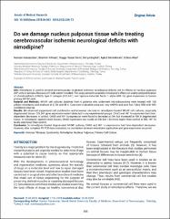
| dc.description.abstract | Aim: Nimodipine is used to prevent cerebrovascular-originated ischemic neurological deficits, yet its effects on nucleus pulposus (NP) cells or annulus fibrosus (AF) cells weren’t studied. This study aimed to examine nimodipine’s effects on vitality and proliferation of chondroadherin (CHAD), type II collagen (COL2A1), and hypoxia-inducible factor 1 alpha (HIF 1?) gene expression in human primary NP/AF cells. Material and Methods: NP/AF cell cultures obtained from 6 patients who underwent microdiscectomy were treated with 100 µMolar nimodipine and analyzed at 0, 24, and 48 h. Data were evaluated using one-way ANOVA and post-hoc Tukey HSD with 95% confidence interval. Results: We observed suppressed cell proliferation and increased necrosis in nimodipine-treated NP/AF cell cultures, especially degenerated tissue. COL2A1 gene expression wasn’t detected in any experimental groups. CHAD and HIF 1? expression had timedependent decreases in control. CHAD and HIF 1? expression were found to decrease at 24h, but increased at 48h in degenerated tissue. In nimodipine-applied intact tissues, CHAD expression was stable at 24h but 1.62 times higher than control at 48h. HIF 1? levels were lower than control. Conclusion: In nimodipine-treated degenerated AF/NP cultures, CHAD and HIF 1? expressions had time-dependent decreases. However, after complete RT-PCR data evaluation, no correlation between nimodipine application and gene expression occurred. | en_US |
| dc.language.iso | eng | en_US |
| dc.rights | info:eu-repo/semantics/openAccess | en_US |
| dc.subject | Annulus Fibrosus | en_US |
| dc.subject | Cytotoxicity | en_US |
| dc.subject | Nimodipine | en_US |
| dc.subject | Nucleus Pulposus | en_US |
| dc.subject | Primary Cell Culture | en_US |
| dc.title | Do we damage nucleus pulposus tissue while treating cerebrovascular ischemic neurological deficits with nimodipine? | en_US |
| dc.type | article | en_US |
| dc.relation.ispartof | Annals of Medical Research | en_US |
| dc.department | İstanbul Medipol Üniversitesi, Tıp Fakültesi, Dahili Tıp Bilimleri Bölümü, Tıbbi Farmakoloji Ana Bilim Dalı | en_US |
| dc.authorid | 0000-0003-2003-6337 | en_US |
| dc.identifier.volume | 25 | en_US |
| dc.identifier.issue | 2 | en_US |
| dc.identifier.startpage | 266 | en_US |
| dc.identifier.endpage | 273 | en_US |
| dc.relation.publicationcategory | Makale - Ulusal Hakemli Dergi - Kurum Öğretim Elemanı | en_US |


