| dc.contributor.author | Kanat, Mustafa | |
| dc.contributor.author | Vardı, Şeref | |
| dc.contributor.author | Arınç, Hüseyin | |
| dc.contributor.author | Gündüz, Hüseyin | |
| dc.contributor.author | Erdem, Alim | |
| dc.contributor.author | Karagöz, Yalçın | |
| dc.date.accessioned | 10.07.201910:49:13 | |
| dc.date.accessioned | 2019-07-10T20:01:37Z | |
| dc.date.available | 10.07.201910:49:13 | |
| dc.date.available | 2019-07-10T20:01:37Z | |
| dc.date.issued | 2013 | en_US |
| dc.identifier.citation | Kanat, M., Vardı, Ş., Arınç, H., Gündüz, H., Erdem, A. ve Karagöz, Y. (2013). Evaluation of cardiac functions with tissue doppler ımaging in prediabetic subjects. Korean Circulation Journal, 43(2), 82-86. https://dx.doi.org/10.4070/kcj.2013.43.2.82 | en_US |
| dc.identifier.issn | 1738-5520 | |
| dc.identifier.issn | 1738-5555 | |
| dc.identifier.uri | https://dx.doi.org/10.4070/kcj.2013.43.2.82 | |
| dc.identifier.uri | https://hdl.handle.net/20.500.12511/3368 | |
| dc.description | WOS: 000342383800003 | en_US |
| dc.description | PubMed ID: 23508684 | en_US |
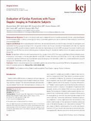
| dc.description.abstract | Background and Objectives: The aim of the present study was to evaluate left ventricle systolic and diastolic function, using tissue Doppler echocardiography (TDE), in relation to blood glucose status in prediabetic patients who had no evidence of heart disease by conventional echocardiography (CE). Subjects and Methods: We included 60 patients (30 female, 30 male) and 20 healthy controls (10 male, 10 female). All participants were randomised into four groups according to their oral glucose tolerance test. Group-I consisted of those patients who had only impaired fasting glucose (IFG). group-II consisted of patients who had only impaired glucose tolerance (IGT) and group-III consisted of patients who had both IFG and IGT, that is so-called combined glucose intolerance. Group-IV included the healthy controls. All subjects underwent both CE and TDE. Results: No significant differences were found among the four groups in terms of CE. There was no significant difference between group-IV and group-I with respect to the early peak diastolic velocity (Ea) of medial mitral annulus (11.65 +/- 0.66 vs. 9.72 +/- 1.58, p>0.05), whereas a statistically significant difference was found between group-IV and group-II (11.65 +/- 0.66 vs. 9.06 +/- 1.07, p<0.001) and between group-IV and group-III (11.65 +/- 0.66 vs. 9.74 +/- 1.09, p<0.05). Conclusion: Diastolic myocardial dysfunction in prediabetic patients may be identified by quantitative TDE before the appearance of CE indices of myocardial dysfunction. | en_US |
| dc.language.iso | eng | en_US |
| dc.publisher | Korean Soc Cardiology | en_US |
| dc.rights | info:eu-repo/semantics/openAccess | en_US |
| dc.subject | Type 2 Diabetes Mellitus | en_US |
| dc.subject | Diabetic Cardiomyopathies | en_US |
| dc.subject | Tissue Doppler Imaging | en_US |
| dc.subject | Glucose Intolerance | en_US |
| dc.title | Evaluation of cardiac functions with tissue doppler ımaging in prediabetic subjects | en_US |
| dc.type | article | en_US |
| dc.relation.ispartof | Korean Circulation Journal | en_US |
| dc.department | İstanbul Medipol Üniversitesi, Tıp Fakültesi, Dahili Tıp Bilimleri Bölümü, İç Hastalıkları Ana Bilim Dalı | en_US |
| dc.identifier.volume | 43 | en_US |
| dc.identifier.issue | 2 | en_US |
| dc.identifier.startpage | 82 | en_US |
| dc.identifier.endpage | 86 | en_US |
| dc.relation.publicationcategory | Makale - Uluslararası Hakemli Dergi - Kurum Öğretim Elemanı | en_US |
| dc.identifier.doi | 10.4070/kcj.2013.43.2.82 | en_US |
| dc.identifier.wosquality | Q2 | en_US |
| dc.identifier.scopusquality | Q2 | en_US |