| dc.contributor.author | Sönmez, Mesut Mehmet | |
| dc.contributor.author | Çamur, Savas | |
| dc.contributor.author | Ertürer, Erden | |
| dc.contributor.author | Uğurlar, Meriç | |
| dc.contributor.author | Kara, Adnan | |
| dc.contributor.author | Öztürk, İrfan | |
| dc.date.accessioned | 10.07.201910:49:13 | |
| dc.date.accessioned | 2019-07-10T20:01:31Z | |
| dc.date.available | 10.07.201910:49:13 | |
| dc.date.available | 2019-07-10T20:01:31Z | |
| dc.date.issued | 2017 | en_US |
| dc.identifier.citation | Sönmez, M. M., Çamuri S., Ertürer, E., Uğurlar, M., Kara, A. ve Öztürk, İ. (2017). Strategies for proximal femoral nailing of unstable intertrochanteric fractures: Lateral decubitus position or traction table. Journal of the American Academy of Orthopaedic Surgeons, 25(3), E37-E44. https://dx.doi.org/10.5435/JAAOS-D-15-00691 | en_US |
| dc.identifier.issn | 1067-151X | |
| dc.identifier.issn | 1940-5480 | |
| dc.identifier.uri | https://dx.doi.org/10.5435/JAAOS-D-15-00691 | |
| dc.identifier.uri | https://hdl.handle.net/20.500.12511/3318 | |
| dc.description | WOS: 000404858500001 | en_US |
| dc.description | PubMed ID: 28134676 | en_US |
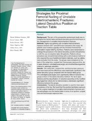
| dc.description.abstract | Background: The aim of this prospective randomized study was to compare the traction table and lateral decubitus position techniques in the management of unstable intertrochanteric fractures. Methods: Eighty-two patients with unstable intertrochanteric fractures between 2011 and 2013 were included in this study. All patients were treated surgically with the Proximal Femoral Nail Antirotation implant (DePuy Synthes). Patients were randomized to undergo the procedure in the lateral decubitus position (42 patients) or with the use of a traction table (40 patients). Patients whose procedure was not performed entirely with a semi-invasive method or who required the use of additional fixation materials, such as cables, were excluded from the study. The groups were compared on the basis of the setup time, surgical time, fluoroscopic exposure time, tip-to-apex distance, collodiaphyseal angle, and modified Baumgaertner criteria for radiologic reduction. Results: The setup time, surgical time, and fluoroscopic exposure time were lower and the differences were statistically significant in the lateral decubitus group compared with the traction table group. The collodiaphyseal angles were significantly different between the groups in favor of the lateral decubitus method. The tip-to-apex distance and the classification of reduction according to the modified Baumgaertner criteria did not demonstrate a statistically significant difference between the groups. Conclusions: The lateral decubitus position is used for most open procedures of the hip. We found that this position facilitates exposure for the surgical treatment of unstable intertrochanteric fractures and has advantages over the traction table in terms of set up time, surgical time and fluoroscopic exposure time. | en_US |
| dc.language.iso | eng | en_US |
| dc.publisher | Lippincott Williams & Wilkins | en_US |
| dc.rights | info:eu-repo/semantics/embargoedAccess | en_US |
| dc.subject | Strategies for Proximal Femoral Nailing | en_US |
| dc.subject | Intertrochanteric Fractures | en_US |
| dc.subject | Lateral Decubitus Position | en_US |
| dc.subject | Traction Table | en_US |
| dc.title | Strategies for proximal femoral nailing of unstable intertrochanteric fractures: Lateral decubitus position or traction table | en_US |
| dc.type | article | en_US |
| dc.relation.ispartof | Journal of the American Academy of Orthopaedic Surgeons | en_US |
| dc.department | İstanbul Medipol Üniversitesi, Tıp Fakültesi, Cerrahi Tıp Bilimleri Bölümü, Ortopedi ve Travmatoloji Ana Bilim Dalı | en_US |
| dc.authorid | 0000-0001-8437-5405 | en_US |
| dc.identifier.volume | 25 | en_US |
| dc.identifier.issue | 3 | en_US |
| dc.identifier.startpage | E37 | en_US |
| dc.identifier.endpage | E44 | en_US |
| dc.relation.publicationcategory | Makale - Uluslararası Hakemli Dergi - Kurum Öğretim Elemanı | en_US |
| dc.identifier.doi | 10.5435/JAAOS-D-15-00691 | en_US |
| dc.identifier.wosquality | Q2 | en_US |
| dc.identifier.scopusquality | Q1 | en_US |


