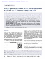
Immunocytoexpression profile of ProExC in smears interpreted as ASC-US, ASC-H, and cervical intraepithelial lesion
| dc.contributor.author | Tosuner, Zeynep | |
| dc.contributor.author | Türkmen, İlknur | |
| dc.contributor.author | Arıcı, Sema | |
| dc.contributor.author | Sönmez, Cavide | |
| dc.contributor.author | Turna, Seval | |
| dc.contributor.author | Onaran, Öykü | |
| dc.date.accessioned | 10.07.201910:49:13 | |
| dc.date.accessioned | 2019-07-10T19:57:57Z | |
| dc.date.available | 10.07.201910:49:13 | |
| dc.date.available | 2019-07-10T19:57:57Z | |
| dc.date.issued | 2017 | en_US |
| dc.identifier.citation | Tosuner, Z., Türkmen, İ., Arıcı, S., Sönmez, C., Turna, S. ve Onaran, Ö. (2017). Immunocytoexpression profile of ProExC in smears interpreted as ASC-US, ASC-H, and cervical intraepithelial lesion. Journal of Cytology, 34(1), 34-38. https://dx.doi.org/10.4103/0970-9371.197605 | en_US |
| dc.identifier.issn | 0970-9371 | |
| dc.identifier.issn | 0974-5165 | |
| dc.identifier.uri | https://dx.doi.org/10.4103/0970-9371.197605 | |
| dc.identifier.uri | https://hdl.handle.net/20.500.12511/3080 | |
| dc.description | WOS: 000393891700007 | en_US |
| dc.description | PubMed ID: 28182079 | en_US |
| dc.description.abstract | Aims: We aimed to investigate the immunocytoexpression profiles of a novel assay ProEx C for topoisomerase II alpha (TOP2A) and minichromosome maintenance protein 2 (MCM2) in abnormal interpreted smears. Settings and Design: Screening programs with Papanicolaou smear and high risk group human papilloma virus testing have yielded a dramatic reduction of cervical cancer incidence. However, both of these tests have limited specificity for the detection of clinically significant cervical high grade lesions. ProEx C for topoisomerase II alpha (TOP2A) and minichromosome maintenance protein 2 (MCM2) has been considered to have tight association with high grade intraepithelial lesions. Materials and Methods: A total number of 54 SurePath cervical cytology specimens of patients previously interpreted as atypical squamous cells-undetermined significance (ASC-US), atypical squamous cells-cannot exclude high grade squamous intraepithelial lesion (ASC-H), low grade squamous intraepithelial lesion (LSIL), and high grade squamous intraepithelial lesion (HSIL) were included in our study. Results and Conclusions: ProEx C was positive in 14 of HSILs (100%), 3 of 19 LSILs (16%), 2 of 4 ASC-Hs, and none of ASC-USs (0%). The ProEx C test showed very intense nuclear staining in all cytologically abnormal cells. Further studies are indicated to evaluate the diagnostic role of ProEx C. | en_US |
| dc.language.iso | eng | en_US |
| dc.publisher | Medknow Publications & Media Pvt Ltd | en_US |
| dc.rights | info:eu-repo/semantics/openAccess | en_US |
| dc.rights | Attribution-NonCommercial-ShareAlike 3.0 Unported | * |
| dc.rights.uri | https://creativecommons.org/licenses/by-nc-sa/3.0/ | * |
| dc.subject | ASC-H | en_US |
| dc.subject | ASC-US | en_US |
| dc.subject | HSIL | en_US |
| dc.subject | Immunocytochemistry | en_US |
| dc.subject | LSIL | en_US |
| dc.subject | ProEx C | en_US |
| dc.title | Immunocytoexpression profile of ProExC in smears interpreted as ASC-US, ASC-H, and cervical intraepithelial lesion | en_US |
| dc.type | article | en_US |
| dc.relation.ispartof | Journal of Cytology | en_US |
| dc.department | İstanbul Medipol Üniversitesi, Tıp Fakültesi, Cerrahi Tıp Bilimleri Bölümü, Tıbbi Patoloji Ana Bilim Dalı | en_US |
| dc.identifier.volume | 34 | en_US |
| dc.identifier.issue | 1 | en_US |
| dc.identifier.startpage | 34 | en_US |
| dc.identifier.endpage | 38 | en_US |
| dc.relation.publicationcategory | Makale - Uluslararası Hakemli Dergi - Kurum Öğretim Elemanı | en_US |
| dc.identifier.doi | 10.4103/0970-9371.197605 | en_US |
| dc.identifier.wosquality | Q4 | en_US |
| dc.identifier.scopusquality | Q3 | en_US |
Bu öğenin dosyaları:
Bu öğe aşağıdaki koleksiyon(lar)da görünmektedir.
-
Makale Koleksiyonu [3756]
Article Collection -
PubMed İndeksli Yayınlar Koleksiyonu [4210]
PubMed Indexed Publications Collection -
Scopus İndeksli Yayınlar Koleksiyonu [6535]
Scopus Indexed Publications Collection -
WoS İndeksli Yayınlar Koleksiyonu [6598]
WoS Indexed Publications Collection

Aksi belirtilmediği sürece bu öğenin lisansı: info:eu-repo/semantics/openAccess


