| dc.contributor.author | Gürler, Gökhan | |
| dc.contributor.author | Delilbaşı, Çağrı | |
| dc.date.accessioned | 10.07.201910:49:13 | |
| dc.date.accessioned | 2019-07-10T19:56:46Z | |
| dc.date.available | 10.07.201910:49:13 | |
| dc.date.available | 2019-07-10T19:56:46Z | |
| dc.date.issued | 2015 | en_US |
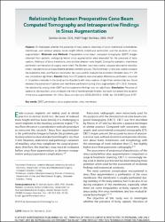
| dc.identifier.citation | Gürler, G. ve Delilbaşı, Ç. (2015). Relationship between preoperative cone beam computed tomography and intraoperative findings in sinus augmentation. International Journal of Oral and Maxillofacial Implants, 30(6), 1244-1248. https://dx.doi.org/10.11607/jomi.3797 | en_US |
| dc.identifier.issn | 0882-2786 | |
| dc.identifier.issn | 1942-4434 | |
| dc.identifier.uri | https://dx.doi.org/10.11607/jomi.3797 | |
| dc.identifier.uri | https://hdl.handle.net/20.500.12511/2809 | |
| dc.description | WOS: 000367254100002 | en_US |
| dc.description | PubMed ID: 26574849 | en_US |
| dc.description.abstract | Purpose: To investigate whether the presence of bony septum, thickness of sinus membrane (schneiderian membrane), and residual alveolar bone height affects membrane perforation and the duration of sinus augmentation. Materials and Methods: Preoperative cone beam computed tomography (CBCT) images obtained from patients undergoing lateral sinus augmentation were evaluated for the presence of bony septum, thickness of sinus membrane, and residual alveolar bone height. During the operation, membrane perforation and duration of surgery were noted. The Student t test was used to compare descriptive statistics (mean, standard error) and quantitative variables between groups. The Fisher exact. chi(2) test was used to compare the qualitative data, and Pearson correlation test was used to evaluate the correlation between data. P < .05 was considered significant. Results: Data from 57 patients were evaluated. Membrane perforation occurred in 14 patients included in the study and in 8 patients with sinus septum. A significant relationship was found between the presence of septum and membrane perforation during sinus augmentation (P = .014). However, the relationship among other CBCT and intraoperative findings was not significant. Conclusion: Presence of septum in the maxillary sinus increases the risk of membrane perforation, but does not extend the duration of the sinus augmentation. | en_US |
| dc.language.iso | eng | en_US |
| dc.publisher | Quintessence Publishing | en_US |
| dc.rights | info:eu-repo/semantics/openAccess | en_US |
| dc.subject | CBCT | en_US |
| dc.subject | Perforation | en_US |
| dc.subject | Sinus Augmentation | en_US |
| dc.subject | Sinus Membrane | en_US |
| dc.title | Relationship between preoperative cone beam computed tomography and intraoperative findings in sinus augmentation | en_US |
| dc.type | article | en_US |
| dc.relation.ispartof | International Journal of Oral and Maxillofacial Implants | en_US |
| dc.department | İstanbul Medipol Üniversitesi, Diş Hekimliği Fakültesi, Ağız, Diş ve Çene Cerrahisi Ana Bilim Dalı | en_US |
| dc.authorid | 0000-0002-6705-3110 | en_US |
| dc.authorid | 0000-0003-3347-1151 | en_US |
| dc.identifier.volume | 30 | en_US |
| dc.identifier.issue | 6 | en_US |
| dc.identifier.startpage | 1244 | en_US |
| dc.identifier.endpage | 1248 | en_US |
| dc.relation.publicationcategory | Makale - Uluslararası Hakemli Dergi - Kurum Öğretim Elemanı | en_US |
| dc.identifier.doi | 10.11607/jomi.3797 | en_US |
| dc.identifier.wosquality | Q2 | en_US |
| dc.identifier.scopusquality | Q1 | en_US |