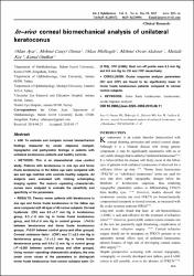
In-vivo corneal biomechanical analysis of unilateral keratoconus
Künye
Ayar, O., Özmen, M. C., Müftüoǧlu, O., Akdemir, M. O., Koç, M. ve Özülken, K. (2015). In-vivo corneal biomechanical analysis of unilateral keratoconus. International Journal of Ophthalmology, 8(6), 1141-1145. https://dx.doi.org/10.3980/j.issn.2222-3959.2015.06.11Özet
AIM: To evaluate and compare corneal biomechanical findings measured by ocular response analyzer, topographic and pachymetric findings in patients with unilateral keratoconus patients and healthy controls. METHODS: This is an observational, case -control study. Patients with keratoconus in one eye and forme fruste keratoconus in the fellow eye were compared with sex and age matched with controls healthy subjects. All subjects were evaluated with rotating scheimpflug imaging system. The receiver -operating -characteristic curves were analyzed to evaluate the sensitivity and specificity of the parameters. RESULTS: Twenty-seven patients with keratoconus in one eye and forme fruste keratoconus in the fellow eye were compared with 40 eyes of 40 normal subjects. Corneal hysteresis (CH) was 8.0 +/- 1.7 mm Hg in keratoconus group, 8.3 +/- 1.6 mm Hg in forme fruste keratoconus group, and 9.8 +/- 1.6 mm Hg in control groups (P=0.54 between keratoconus and forme fruste keratoconus groups, P<0.01 between control group and other groups). Corneal resistance factor (CRF) was 7.1 +/- 2.2 mm Hg in keratoconus group, 7.8 +/- 1.2 mm Hg in forme fruste keratoconus group and 9.9 +/- 1.5 mm Hg in control group (P < 0.001 between control group and other groups). Using receiver-operating-characteristic analysis, the area under curve values of the parameters to distinguish forme fruste keratoconus from control subjects were: CH (0.768), CRF (0.866). Best cut-off points were 9.3 mm Hg and 8.8 mm Hg for CH and CRF respectively. CONCLUSION: Ocular response analyzer parameters (CH and CRF) are found to be significantly lower in forme fruste keratoconus patients compared to normal control subjects.
WoS Q Kategorisi
Q4Scopus Q Kategorisi
Q3Kaynak
International Journal of OphthalmologyCilt
8Sayı
6Bağlantı
https://dx.doi.org/10.3980/j.issn.2222-3959.2015.06.11https://hdl.handle.net/20.500.12511/2799
Koleksiyonlar
- Makale Koleksiyonu [3751]
- PubMed İndeksli Yayınlar Koleksiyonu [4203]
- Scopus İndeksli Yayınlar Koleksiyonu [6522]
- WoS İndeksli Yayınlar Koleksiyonu [6585]


