| dc.contributor.author | Durur Subaşı, Irmak | |
| dc.contributor.author | Durur Karakaya, Afak | |
| dc.contributor.author | Karaman, Adem | |
| dc.contributor.author | Şeker, Mehmet | |
| dc.contributor.author | Demirci, Elif | |
| dc.contributor.author | Alper, Fatih | |
| dc.date.accessioned | 10.07.201910:49:13 | |
| dc.date.accessioned | 2019-07-10T19:50:56Z | |
| dc.date.available | 10.07.201910:49:13 | |
| dc.date.available | 2019-07-10T19:50:56Z | |
| dc.date.issued | 2017 | en_US |
| dc.identifier.citation | Durur Subaşı, I., Durur Karakaya, A., Karaman, A., Şeker, M., Demirci, E. ve Alper, F. (2017). Is the necrosis/wall ADC ratio useful for the differentiation of benign and malignant breast lesions? British Journal of Radiology, 90(1073). https://dx.doi.org/10.1259/bjr.20160803 | en_US |
| dc.identifier.issn | 0007-1285 | |
| dc.identifier.issn | 1748-880X | |
| dc.identifier.uri | https://dx.doi.org/10.1259/bjr.20160803 | |
| dc.identifier.uri | https://hdl.handle.net/20.500.12511/2111 | |
| dc.description | WOS: 000402802300017 | en_US |
| dc.description | PubMed ID: 28339285 | en_US |
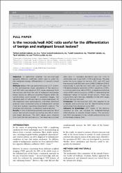
| dc.description.abstract | Objective: To determine whether the necrosis/wall apparent diffusion coefficient (ADC) ratio is useful for the malignant-benign differentiation of necrotic breast lesions. Methods: Breast MRI was performed using a 3-T system. In this retrospective study, calculation of the necrosis/wall ADC ratio was based on ADC values measured from the necrosis and from the wall of malignant and benign breast lesions by diffusion-weighted imaging (DWI). By synchronizing post-contrast T1 weighted images, the separate parts of wall and necrosis were maintained. All the diagnoses were pathologically confirmed. Statistical analyses were conducted using an independent sample t-test and receiver operating characteristic analysis. The intraclass and interclass correlations were evaluated. Results: A total of 66 female patients were enrolled, 38 of whom had necrotic breast carcinomas and 28 of whom had breast abscesses. The ADC values were obtained from both the wall and necrosis. The mean necrosis/wall ADC ratio (6 standard deviation) was 1.6160.51 in carcinomas, and it was 0.6560.33 in abscesses. The area under the curve values for necrosis ADC, wall ADC and the necrosis/wall ADC ratio were 0.680, 0.068 and 0.942, respectively. A wall/necrosis ADC ratio cut-off value of 1.18 demonstrated a sensitivity of 97%, specificity of 93%, a positive-predictive value of 95%, a negative-predictive value of 96% and an accuracy of 95% in determining the malignant nature of necrotic breast lesions. There was a good intra- and interclass reliability for the ADC values of both necrosis and wall. Conclusion: The necrosis/wall ADC ratio appears to be a reliable and promising tool for discriminating breast carcinomas from abscesses using DWI. Advances in knowledge: ADC values of the necrosis obtained by DWI are valuable for malignant-benign differentiation in necrotic breast lesions. The necrosis/wall ADC ratio appears to be a reliable and promising tool in the breast imaging field. | en_US |
| dc.language.iso | eng | en_US |
| dc.publisher | British Institute of Radiology | en_US |
| dc.rights | info:eu-repo/semantics/openAccess | en_US |
| dc.subject | ADC Ratio | en_US |
| dc.subject | Malignant Breast Lesions | en_US |
| dc.subject | Differentiation of Benign | en_US |
| dc.title | Is the necrosis/wall ADC ratio useful for the differentiation of benign and malignant breast lesions? | en_US |
| dc.type | article | en_US |
| dc.relation.ispartof | British Journal of Radiology | en_US |
| dc.department | İstanbul Medipol Üniversitesi, Tıp Fakültesi, Dahili Tıp Bilimleri Bölümü, Radyoloji Ana Bilim Dalı | en_US |
| dc.authorid | 0000-0002-6745-0159 | en_US |
| dc.identifier.volume | 90 | en_US |
| dc.identifier.issue | 1073 | en_US |
| dc.relation.publicationcategory | Makale - Uluslararası Hakemli Dergi - Kurum Öğretim Elemanı | en_US |
| dc.identifier.doi | 10.1259/bjr.20160803 | en_US |
| dc.identifier.wosquality | Q2 | en_US |
| dc.identifier.scopusquality | Q2 | en_US |