• Türkçe
    • English
  • English 
    • Türkçe
    • English
  • Login
View Item 
  •   DSpace@Medipol
  • Araştırma Çıktıları | TR-Dizin | WoS | Scopus | PubMed
  • Scopus İndeksli Yayınlar Koleksiyonu
  • View Item
  •   DSpace@Medipol
  • Araştırma Çıktıları | TR-Dizin | WoS | Scopus | PubMed
  • Scopus İndeksli Yayınlar Koleksiyonu
  • View Item
JavaScript is disabled for your browser. Some features of this site may not work without it.

DeepMQ: A deep learning approach based myelin quantification in microscopic fluorescence images
Thumbnail

View/Open

Tam Metin / Full Text (9.038Mb)

Access

info:eu-repo/semantics/openAccess

Date

2018

Author

Çimen, Sibel
Çapar, Abdülkerim
Ekinci, Dursun Ali
Ayten, Umut Engin
Kerman, Bilal Ersen
Töreyin, Behçet Uğur

Metadata

Show full item record

Citation

Çimen, S., Çapar, A., Ekinci, D. A., Ayten, U. E., Kerman, B. E. ve Töreyin, B. U. (2018). DeepMQ: A deep learning approach based myelin quantification in microscopic fluorescence images. European Signal Processing Conference (EUSIPCO) içinde (61-65. ss.). Rome, Italy, 3-7 September, 2018. https://dx.doi.org/10.23919/EUSIPCO.2018.8553438

Abstract

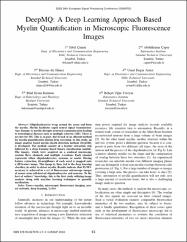
Oligodendrocytes wrap around the axons and form the myelin. Myelin facilitates rapid neural signal transmission. Any damage to myelin disrupts neuronal communication leading to neurological diseases such as multiple sclerosis (MS). There is no cure for MS. This is, in part, due to lack of an efficient method for myelin quantification during drug screening. In this study, an image analysis based myelin sheath detection method, DeepMQ, is developed. The method consists of a feature extraction step followed by a deep learning based binary classification module. The images, which were acquired on a confocal microscope contain three channels and multiple z-sections. Each channel represents either oligodendroyctes, neurons, or nuclei. During feature extraction, 26-neighbours of each voxel is mapped onto a 2D feature image. This image is, then, fed to the deep learning classifier, in order to detect myelin. Results indicate that 93.38% accuracy is achieved in a set of fluorescence microscope images of mouse stem cell-derived oligodendroyctes and neurons. To the best of authors' knowledge, this is the first study utilizing image analysis along with machine learning techniques to quantify myelination.

Source

European Signal Processing Conference (EUSIPCO)

URI

https://hdl.handle.net/20.500.12511/1348
https://dx.doi.org/10.23919/EUSIPCO.2018.8553438

Collections

  • Bildiri Koleksiyonu [56]
  • Scopus İndeksli Yayınlar Koleksiyonu [5808]
  • WoS İndeksli Yayınlar Koleksiyonu [5978]


DSpace software copyright © 2002-2015  DuraSpace
Contact Us | Send Feedback
Theme by 
@mire NV


| Guide | Contact |

DSpace@Medipol

by OpenAIRE
Advanced Search

sherpa/romeo

Browse

All of DSpaceCommunities & CollectionsBy Issue DateAuthorsInstitution AuthorORCIDTitlesSubjectsTypeLanguageDepartmentCategoryWoS Q ValueScopus Q ValuePublisherAccess TypeThis CollectionBy Issue DateAuthorsInstitution AuthorORCIDTitlesSubjectsTypeLanguageDepartmentCategoryWoS Q ValueScopus Q ValuePublisherAccess Type

My Account

LoginRegister

Statistics

View Google Analytics Statistics

DSpace software copyright © 2002-2015  DuraSpace
Contact Us | Send Feedback
Theme by 
@mire NV


|| Guide || Library || İstanbul Medipol University || OAI-PMH ||

Kütüphane ve Dokümantasyon Daire Başkanlığı, İstabul, Turkey
If you find any errors in content, please contact: [email protected]

Creative Commons License
DSpace@Medipol by İstanbul Medipol University Institutional Repository is licensed under a Creative Commons Attribution-NonCommercial-NoDerivs 4.0 Unported License..

DSpace@Medipol:


DSpace 6.2

tarafından İdeal DSpace hizmetleri çerçevesinde özelleştirilerek kurulmuştur.