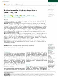
Retinal vascular findings in patients with COVID-19
| dc.contributor.author | Aydemir, Emre | |
| dc.contributor.author | Bayat, Alper Halil | |
| dc.contributor.author | Ören, Burak | |
| dc.contributor.author | Ateşoğlu, Halil İbrahim | |
| dc.contributor.author | Göker, Yasin Şakir | |
| dc.contributor.author | Özçelik, Kazım Çağlar | |
| dc.date.accessioned | 2021-09-06T11:32:02Z | |
| dc.date.available | 2021-09-06T11:32:02Z | |
| dc.date.issued | 2021 | en_US |
| dc.identifier.citation | Aydemir, E., Bayat, A. H., Ören, B., Ateşoğlu, H. İ., Göker, Y. Ş. ve Özçelik, K. Ç. (2021). Retinal vascular findings in patients with COVID-19. Therapeutic Advances in Ophthalmology, 13. https://dx.doi.org/10.1177/25158414211030419 | en_US |
| dc.identifier.issn | 2515-8414 | |
| dc.identifier.uri | https://dx.doi.org/10.1177/25158414211030419 | |
| dc.identifier.uri | https://hdl.handle.net/20.500.12511/8009 | |
| dc.description.abstract | Purpose: The purpose of this study was to compare the retinal vascular caliber of COVID-19 patients with that of healthy subjects. Methods: This was a prospective case-control study. Forty-six patients who had COVID-19 were successfully treated, and 38 age- and gender-matched healthy subjects were enrolled in this study. Fundus photography was taken using fundus fluorescein angiography (FA; Visucam 500; Carl Zeiss Meditec, Jena, Germany). Retinal vascular caliber was analyzed with IVAN, a semi-automated retinal vascular analyzer (Nicole J. Ferrier, College of Engineering, Fundus Photography Reading Center, University of Wisconsin, Madison, WI, USA). Central retinal artery equivalent (CRAE), central retinal vein equivalent (CRVE), and artery-vein ratio (AVR) were compared between groups. Results: The mean age was 37.8 +/- 9.5 years in the COVID-19 group (n = 46) and 40 +/- 8 years in the control group (n = 38) (p = 0.45). The mean CRAE was 181.56 +/- 6.40 in the COVID-19 group and 171.29 +/- 15.06 in the control group (p = 0.006). The mean CRVE was 226.34 +/- 23.83 in the COVID-19 group and 210.94 +/- 22.22 in the control group (p = 0.044). AVR was 0.81 +/- 0.09 in the COVID-19 group and 0.82 +/- 0.13 in the control group (p = 0.712). Conclusion: Patients who had COVID-19 have vasodilation in the retinal vascular structure after recovery. As they may be at risk of retinal vascular disease, COVID-19 patients must be followed after recovery. | en_US |
| dc.language.iso | eng | en_US |
| dc.publisher | SAGE Publications Ltd | en_US |
| dc.rights | info:eu-repo/semantics/openAccess | en_US |
| dc.rights | Attribution-NonCommercial 4.0 International | * |
| dc.rights.uri | https://creativecommons.org/licenses/by-nc/4.0/ | * |
| dc.subject | COVID-19 | en_US |
| dc.subject | Retinal Vascular Caliber | en_US |
| dc.subject | Retinal Vasodilation | en_US |
| dc.title | Retinal vascular findings in patients with COVID-19 | en_US |
| dc.type | article | en_US |
| dc.relation.ispartof | Therapeutic Advances in Ophthalmology | en_US |
| dc.department | İstanbul Medipol Üniversitesi, Tıp Fakültesi, Cerrahi Tıp Bilimleri Bölümü, Göz Hastalıkları Ana Bilim Dalı | en_US |
| dc.authorid | 0000-0003-1827-968X | en_US |
| dc.identifier.volume | 13 | en_US |
| dc.relation.publicationcategory | Makale - Uluslararası Hakemli Dergi - Kurum Öğretim Elemanı | en_US |
| dc.identifier.doi | 10.1177/25158414211030419 | en_US |
Bu öğenin dosyaları:
Bu öğe aşağıdaki koleksiyon(lar)da görünmektedir.
-
Makale Koleksiyonu [3701]
Article Collection -
PubMed İndeksli Yayınlar Koleksiyonu [4120]
PubMed Indexed Publications Collection -
WoS İndeksli Yayınlar Koleksiyonu [6516]
WoS Indexed Publications Collection

Aksi belirtilmediği sürece bu öğenin lisansı: info:eu-repo/semantics/openAccess